



Contents
Scroll to:
https://doi.org/10.30895/2312-7821-2025-498
Scroll to:
INTRODUCTION. Lamotrigine (LTG) is among the most commonly prescribed second-generation antiepileptic drugs due to its low teratogenic risk. However, lamotrigine has pronounced neurotoxic, hepatotoxic, dermatotoxic potential (for genetic and metabolic causes) and in some cases can even cause multi-organ failure. Understanding lamotrigine mechanism can help individualise therapy and increase its safety, considering pharmacodynamics and pharmacometabolomics that determine its metabolism, transport, and elimination in a particular patient.
AIM. This study aimed to develop an approach to lamotrigine therapy of epilepsy and other neurological and psychiatric diseases reducing neurotoxicity, with due regard to pharmacogenomics and pharmacometabolomics.
RESULTS. LTG is metabolised in the liver in two pathways: glucuronidation (major pathway) and P-oxidation (minor pathway). As a result, neutral and toxic (reactive) lamotrigine metabolites are produced that can circulate in blood serum for a long time, penetrate the damaged blood-brain barrier in patients with therapy-resistant seizures and have a neurotoxic effect, triggering or maintaining neurotransmission disorders, impaired synaptic plasticity, neuronal apoptosis and other neurodegeneration mechanisms. An important role in lamotrigine neurotoxicity belongs to transport proteins involved in the efflux (excretion) of reactive (toxic) metabolites from the brain into the systemic circulation, as well as from hepatocytes into the gastrointestinal tract by bile and through the kidneys with urine. Genetically determined delayed efflux through the blood-brain barrier (pharmacogenomics) increases lamotrigine neurotoxic potential.
CONCLUSION. To assess the risk of lamotrigine-induced adverse reactions, together with clinically assessing patient’s condition, it is recommended to: 1) monitor drug distribution (blood, hair, saliva, breast milk); 2) analyse potentially toxic metabolites (blood, saliva, hair); 3) perform pharmacogenetic tests for non-functional polymorphisms of genes encoding key transport proteins and enzymes involved in drug metabolism. Results of pharmacogenetic and pharmacometabolic tests applied in the clinical practice of an epileptologist will allow to manage lamotrigine neurotoxiсity.
Shnayder N.A., Bader V.V., Nasyrova R.F., Kissin M.Ya. Multi-Omics in Predicting Lamotrigine Neurotoxicity: Current Opportunities (Review). Safety and Risk of Pharmacotherapy. 2026;14(1):51-65. https://doi.org/10.30895/2312-7821-2025-498
Lamotrigine (LTG) is a second-generation antiepileptic drug (AED) and mood stabiliser. It is a derivative of phenyltriazine (3,5-diamino-6-(2,3-dichlorophenyl)-1,2,4-triazine) used in adults and children over 12 years as a mono- and/or polytherapy for focal and generalised epileptic seizures, as well as in children over two years of age as a concomitant therapy for refractory focal epilepsy and Lennox-Gastaut syndrome [1–3]. LTG was the first drug after lithium to receive approval by the U.S. Food and Drug Administration (FDA) for maintenance treatment of type I bipolar disorder [3][4]. Evidence suggests that LTG may be clinically effective in neuropathic pain and headaches1 [5].
LTG is rapidly and almost completely absorbed after oral administration, with minimal first-pass metabolism in the liver and absolute bioavailability up to 98%. The LTG maximum plasma concentration occurs within 1.4 to 4.8 hours after oral intake and is directly proportional to the dose ranging from 50 to 400 mg (therapeutic plasma concentration varies from 4 to 10 µg/mL) [6]. Specificity of LTG pharmacokinetics and its low teratogenic potential results in its increased applicationand widespread prescription by neurologists and psychiatrists, including female patients of reproductive age [7][8]. However, during pregnancy, LTG plasma concentration decreases on average by 68% [9], while the clearance increases by 300% compared to pre-pregnancy levels [10]. In pregnant women taking standard LTG doses, therapeutic efficiency in epilepsy or bipolar disorder decreases, creating life-threatening conditions for both the mother and the foetus [11].
Insufficient data on LTG pharmacokinetics during pregnancy impair the dose adjustment [12]. It was shown that in LTG, area under the curve (AUC) significantly decreases during pregnancy, thus the dose recommended in the first, second, and third trimester is 3-, 3-, and 5-fold higher, respectively, than the baseline dose taken before pregnancy. However, considering foetal safety, the maximum recommended single daily dose in the first, second, and third trimester should not exceed 400, 500, and 700 mg, respectively, while the total dose for twice-daily administration should be 300, 400, and 600 mg, respectively [13].
Recent studies have demonstrated that higher LTG doses entering the foetal bloodstream through the placenta or the newborn’s bloodstream through the breast milk can lead to serious adverse drug reactions (ADRs) [14][15]; some ADRs include foetal death, severe hyperbilirubinaemia with bilirubin-induced neurologic dysfunction, and even severe birth asphyxia (at LTG blood levels ≥4.87 mg/L in infants) [16].
An average LTG level in umbilical cord blood is approximately 60% of the maternal serum level at delivery [17]. The risk for both the mother taking LTG and the newborn significantly increases in the postpartum period, since the rate of LTG clearance returns to pre-pregnancy values within the first few weeks after delivery [7][18]. According to Clark et al., LTG blood levels increase most dramatically in the first 1.5 weeks postpartum and then vary between 30% and 640% compared to the second and third trimester. The average LTG plasma concentration during the first 4 weeks postpartum is 154% to 402% higher than in the third trimester.
Pharmacokinetic studies of LTG levels in the blood of pregnant women, foetuses, newborns, and infants [19], as well as in breast milk [20] are essential for toxicity assessment and timely dose adjustment, while being difficult to implement in clinical practice. This review presents promising multi-omics (pharmacogenomic and pharmacometabolomic) methods for precise and safe LTG prescription [21].
The aim of the study is to develop an approach to LTG therapy in epilepsy and other neurological / psychiatric disorders considering pharmacogenomics and pharmacometabolomics in order to reduce the risk of LTG neurotoxicity.
The bibliographic research was performed between March 2010 and March 2025 (Figure 1) according to PRISMA 20202 guidelines in bibliographic databases using inclusion/exclusion criteria, keywords, and their combinations in the Russian and English language (Table 1). Following the exclusion of duplicate publications and negative studies, 35 out of 134 publications meeting the stated objectives and search criteria were included in this review.

The figure was prepared by the authors using their own data
Figure 1. PRISMA flowchart showing the number of publications selected for a review
Table 1. Inclusion and exclusion criteria of publications in the review
|
Parameters |
Inclusion criteria |
Exclusion criteria |
|
Publication type |
Full-text versions: original articles, clinical cases, systematic reviews, meta-analyses, and Cochrane reviews |
Abstracts, conference materials, posters, and dissertations |
|
Access to publication |
Access to the full version |
No access to the full version |
|
Publication language |
English, Russian |
Other languages |
|
Databases |
eLIBRARY.RU, PubMed, Scopus, Springer Nature Link, Clinical Keys, Google Scholar |
Other databases |
|
Internet resource |
State Register of Medicinal Products, U.S. Food and Drug Administration, DrugBank, PharmGKB, Online Mendelian Inheritance in Man, Human Metabolome Database, PubChem |
Other online resources |
|
Search depth |
2010–2025 (15 years) |
Until March 2010 |
|
Keywords |
Lamotrigine; antiepileptic drug; lamotrigine metabolism; glucuronidation; P-oxidation; metabolites; pharmacogenetics; pharmacometabolomics; adverse drug reaction; neurotoxiсity |
Not applicable |
The table was prepared by the authors using their own data
The role of ATP-binding cassette (ABC) transporter superfamily in the LTG efflux (transport from the brain into the bloodstream) involves contradictory data, particularly for P-glycoprotein (P-gp) encoded by ABCB1 gene (MDR1) [6] and breast cancer resistance protein encoded by gene member 2 of the ATP-binding cassette sub-family G (ATP-binding cassette sub-family G member 2, ABCG2; breast cancer resistance protein). Polymorphisms in genes encoding this transporter protein may significantly affect LTG pharmacokinetics and bioavailability [22].
LTG has been identified as a substrate for two main efflux transport proteins through the blood-brain barrier (BBB) – P-glycoprotein (P-gp) and ABCG2 – that play a synergistic or cooperative role in the efflux of dual substrates [23]. These transporter proteins may strongly influence the variable CNS response to LTG, particularly in epileptic patients who overexpress both transporters. Moreover, polymorphisms in ABCC2 (MRP2) gene encoding canalicular multispecific organic anion transporter (multidrug resistance-associated protein 2, MRP2) are associated with drug resistance in different populations; this indicates the transporter involvement in LTG efflux [24]. ABCC3 (MRP3) protein is located on the hepatic sinusoid wall separating the cytosol from the bloodstream and transports LTG glucuronides from the liver cells into the circulation for further renal elimination [25].
The mechanism allowing LTG to cross the BBB is not entirely clear; however, the role of organic cation transporters (OCT) in LTG transport has been identified. Specifically, LTG is a substrate for the transporter protein SLC22A1, also known as OCT1 [27].
LTG shows affinity to several types of receptors expressed in the brain. It produces weak inhibition of serotonin receptors (5-HT2 and 5-HT3), weak binding to adenosine A1/A2 receptors, adrenergic receptors (α1/α2/β), dopamine 1 and 2 receptors (D1/D2), gamma-aminobutyric acid (GABA) A/B receptors, histamine H1 receptors, κ-opioid receptors (KOR), and muscarinic acetylcholine receptors (mACh). LTG also exerts a weak inhibitory effect on sigma-opioid receptors3. However, the leading hypothesis for the LTG anticonvulsant effect is its binding to voltage-gated sodium channels [28], heteromeric complexes regulating sodium exchange between the intra- and extracellular space between neurons.
LTG is believed to selectively inhibit voltage-gated sodium channels type 2 (VGSC) [29] by stabilising the neuronal membrane and thereby inhibiting presynaptic release of excitatory neurotransmitters (glutamate and aspartate). Sodium channel alpha subunits in the human brain have four major isoforms: SCN1A, SCN2A, SCN3A, and SCN8A [29][30]. At rest, a sodium channel pore is closed. When neuronal membranes containing VGSC are sufficiently depolarised during an action potential, the protein changes its structure, causing the pore to open. Within milliseconds after opening, inactivation begins that blocks ion flow through the channel. At this point, the channel is considered deactivated. LTG binds to inactivated VGSC, thus acting as an antagonist [28].
LTG may also interact with N-, P-, and Q-type voltage-gated calcium channels (VGCC) encoded by the CACN gene, which makes LTG a broad-spectrum medicinal product. VGCCs are structurally similar to VGSCs but lack the intracellular inactivation loop typical for VGSCs. LTG inhibits Cav2.3 (R-type) calcium currents, which may represent one of its antiepileptic mechanisms [31].
Although LTG is not considered to directly target GABAergic receptors, they are the primary CNS inhibitory receptors; heterogeneity of GABA ionotropic receptors is linked to epileptogenesis [32]. Glutamate is an excitatory neurotransmitter playing an important role in epileptogenesis. Increased glutamate levels are associated with seizure initiation [33]. LTG reduces glutamate levels in the brain by inhibiting its overexcitation, which also explains LTG antiepileptic and mood-stabilising effects [33][34].
The primary LTG metabolic pathway is glucuronidation catalysed predominantly by enzymes of the uridine 5'-diphospho-glucuronosyltransferase (UGT) system, specifically UGT1A4 and UGT2B7 [35][36]; other involved UGT enzymes are UGT1A3, UGT2B15, UGT1A42, and UGT2B72 [36–38]. These UGT enzymes play a significant role in the interindividual variability of LTG metabolism.
LTG undergoes glucuronidation to form the following metabolites: LTG-2-N-glucuronide (main metabolite, pharmacologically inactive), LTG-5-N-glucuronide, LTG-2-N-methyl, and other minor compounds4 [28][39]. LTG-2-N-glucuronide is a primary metabolite of UGT1A4 enzyme [12]. Valproic acid is known to inhibit LTG metabolism in the liver by increasing its half-life linearly (24 to about 72 hours) alongside with the dose [28]. The increased plasma concentration-to-dose ratio (C/D ratio) results from competitive inhibition of UGT1A4 or UGT2B7 enzymes by valproates [38] (valproic acid has a higher affinity for UGT2B7 and is also a substrate for UGT1A4 [39]), an important consideration in polytherapy.
Two promoter polymorphisms (rs3732218 −163G>A and rs373221 −219C>T) in the UGT1A4 gene are associated with a 40–50% reduction in UGT1A4 enzyme activity and an increased risk of toxic LTG blood concentrations at average therapeutic doses [40]. The 142T>G polymorphism (rs2011425) in the coding region of the UGT1A4 gene is linked to increased LTG blood concentrations and decreased clearance in patients with homozygous TT genotype compared to GT and GG genotypes [38, 41]. The rs2011425 polymorphism (UGT1A4*3) of the UGT1A4 gene was associated with reduced concentration-to-dose (C/D) ratios during pregnancy [42]. Patients with the homozygous rs2011425 GG genotype (extensive or normal metabolisers) exhibit higher glucuronidation activity compared to those with the TT genotype (slow metabolisers) [43]. The rs6755571 polymorphism in the UGT1A4 gene (70C>A) is associated with higher LTG blood concentrations and lower clearance, even during pregnancy [41][42]. The rare rs34946978 polymorphism (more common in East Asians than Caucasians or Africans) is linked to a marked reduction in LTG glucuronidation activity. The impact of various haplotypes on LTG pharmacokinetics remains insufficiently studied.
The minor (rare) homozygous TT genotype of the rs7668258 polymorphism (−161C>T) in the UGT2B7 gene is associated with an increased LTG C/D ratio compared to patients with the major (common) CC genotype [45]. Patients with the TT and CT genotypes have an average LTG clearance about 18% lower than those with the CC genotype [46]. A study of the −372A>G polymorphism in the UGT2B7 gene showed that LTG clearance was 247% higher in the major homozygous GG genotype compared to the minor AA genotype [47]. A clinical case has been reported linking the −372A>G polymorphism of UGT2B7 with dermatotoxicity and multiple organ dysfunction syndrome [48]. The rs7439366 polymorphism (802C>T) of UGT2B7 is also associated with a decreased LTG C/D ratio in pregnant women [41].
Patients with the major homozygous CC genotype of the rs6755571 polymorphism (70 C>T) in the UGT1A4 gene (normal metabolisers) showed a 22% decrease in LTG concentration compared to the heterozygous CT genotype (intermediate metabolisers) [36]. Serum LTG level in patients with the minor homozygous TT rs7668258 (−161 C>T) genotype of the UGT2B7 gene (slow metabolisers) was 1.2 times higher than in patients homozygous for the C allele (normal metabolisers) [36][37].
The UGT2B15*2 polymorphism that causes a G>T substitution in the UGT2B15 gene and results in an amino acid change from aspartic acid (D) to tyrosine (Y) at position 85 of UGT2B15 enzyme is present in about 50% of the Caucasian population and to a lesser extent in Hispanic, African American, Chinese, Japanese, and Korean populations (allele frequencies range from 36% to 49%). This polymorphism is associated with significantly reduced LTG glucuronidation and may be a clinically important predictor of interindividual variability in LTG clearance. Patients with the homozygous TT genotype had serum LTG levels 18% higher than those with GG homozygous genotype [36]. Moreover, concomitant use of other products that inhibit UGT2B15, particularly valproate, may further affect LTG plasma level and clearance and potentially cause LTG toxicity, especially in pregnant women (second and third trimester), neonates, and infants [7]. The role of polymorphisms in the UGT1A3 gene that can potentially influence LTG glucuronidation still remains unclear [37][38], partly due to conflicting data on the role of the isoenzyme encoded by this gene in the formation of the main active LTG-2-N-glucuronide metabolite [49].
To a lesser extent LTG metabolism involves the liver cytochrome P450 system. According to L. Liu et al. [50], isoenzymes CYP2A6 and CYP2D6 are responsible for accumulating LTG oxidation products. CYP2A6 appears to be the main enzyme activating LTG in human liver microsomes [51]. Further formation of active LTG metabolites occurs in keratinocytes [28]. However, these results are inconclusive, since no additional studies have demonstrated LTG metabolism through the liver cytochrome P450 system.
Although PubChem database covers LTG metabolites, their biogenesis and physiological significance are insufficiently studied. This warrants an additional research aimed at understanding their metabolism and the potential roles in cellular and molecular processes.
ABCB1 and ABCG2 transporter proteins play a key role in LTG transport across the BBB [52]. This suggests that polymorphisms in the genes encoding these transporter proteins are associated with functional variations in LTG efflux through the BBB and may contribute to interindividual differences in therapeutic resistance and LTG neurotoxicity [28].
Polymorphisms rs1128503 (1236C>T), rs2032582 (2677G>T/A), and rs1045642 (3435C>T) in the ABCB1 gene affect LTG transport and blood concentration: carriers of the haplotype 1236C-2677G-3435C have higher LTG serum concentrations compared to carriers of the haplotype 1236T-2677G-3435T, followed by haplotype 1236T-2677T-3435C [21].
Polymorphisms rs2231142 and rs3114020 of the ABCG2 gene are associated with serum LTG concentration normalised to body weight [21][23]. A study on the rs2231142 (421C>A) polymorphism of ABCG2 showed that homozygous carriers of the minor AA genotype receiving LTG monotherapy had lower minimum LTG blood concentrations than homozygous carriers of the major CC genotype [53]. Moreover, this polymorphism accounted for 4.8% of the variability in LTG serum fluctuations among Chinese epileptic patients [54].
The rs717620 (−24C>T) polymorphism in the ABCC2 gene is associated with LTG resistance and the risk of neurotoxicity in Caucasians and Asians, likely due to compensatory activation of the ABCB1 transporter [55]. In Chinese epileptic patients carrying the ABCC2 −24C>T / 1249G>A / 3972C>T haplotype, there is a high risk of LTG resistance [55]. However, the −24C>T polymorphism in the ABCC2 gene was not linked to LTG resistance in epileptic patients of other races and ethnic groups, including Han Chinese, Croats, and Austrians [28].
LTG plasma concentration and the rs628031 (1A>G) polymorphism in the SLC22A1 gene that encodes efflux transporter SLC22A1 were shown to be linked. Patients with the minor homozygous GG genotype had significantly lower LTG plasma concentrations compared to the major homozygous AA genotype, indicating association of this polymorphism with slowed efflux of the product from the brain into the blood [54].
ABCC3 and ABCC4 (also known as MRP4) proteins are responsible for transporting LTG metabolites from the liver into the bloodstream5. About 80% of LTG glucuronides are excreted in the urine (primary elimination route), including LTG [ -2-N-glucuronide] (main metabolite, 80–90%), LTG [-5-N-glucuronide], LTG [-N-oxide], and LTG [-N-methyl] (Table 2).
Table 2. Major lamotrigine metabolites
|
Metabolite |
HMDB6 |
PubChem CID7 |
Detected in |
Metabolite toxicity |
|
Glucuronidation |
||||
|
LTG-2-N-glucuronide |
0061103 |
164342 |
Blood, urine, bile |
Inactive (non-toxic/neutral) |
|
LTG-5-N-glucuronide |
– |
71236569 |
Blood |
Inactive (non-toxic/neutral) |
|
N-glucuronide |
– |
– |
Blood, urine, bile |
Inactive (non-toxic/neutral) |
|
P-oxidation |
||||
|
LTG arene oxide |
– |
PA1661703658 |
Bile |
Active (potentially toxic) |
|
LTG-N-oxide |
– |
15089895 |
Urine, bile |
Active (potentially toxic) |
|
Methylation |
||||
|
LTG-N-methyl |
– |
91810661 |
Urine, bile |
Active (potentially toxic) |
The table was prepared by the authors
Note. –, data not available.
Non-functional polymorphisms in ABCC3 are very rare (maximum 4.7%), and their prevalence shows interethnic differences. However, the minor A allele of the −1767G>A polymorphism in the ABCC3 gene is associated with a significantly decreased expression of the ABCC3 transporter protein in the human liver [56]; this may slow down the transport of LTG metabolites from hepatocytes into the blood.
ABCC4 (MRP4) transporter encoded by the ABCC4 gene is expressed in multiple organs and can modify the effect of LTG or its metabolites on cells with various outcomes. ABCC4 is located on the basolateral membrane of the choroid plexus epithelium and on the luminal side of brain capillaries [57], which prevents many xenobiotics, including LTG, from rapidly entering the brain by effluxing them from blood capillaries into plasma. ABCC4 is also found on the basolateral membrane of human hepatocytes [58]. Only a few comprehensive studies (mainly in vitro) have been conducted regarding the influence of ABCC4 liver expression on drug pharmacokinetics, including LTG; thus its role warrants further investigation [59].
According to dbSNP9, 30,185 polymorphisms have been registered for ABCC4; of them, two-thirds are located in intronic sequences. Some of these have direct or indirect clinical significance. ABCC4 is also localised on the apical membrane of renal proximal tubules. Polymorphisms rs1751034 and rs3742106 in the ABCC4 gene play an important role in LTG elimination in the urine, being associated with an increased risk of kidney disease [60]; this can cause higher LTG plasma concentrations and an increased ADR risk including neurotoxicity and hepatotoxicity.
An additional elimination pathway for LTG glucuronides is their excretion from hepatocytes into bile by ABCC2 transporter (also known as MRP2)10. Data on genes encoding key enzymes involved in LTG metabolism and transport are summarised in Table 3, Table 4, and Figure 2.
Table 3. Genes encoding key enzymes of lamotrigine metabolism
|
Gene |
Chromosome location |
Online Mendelian Inheritance in Men11 |
Enzyme |
Organs with the highest expression |
|
UGT1A4 |
2q37.1 |
606429 |
UGT1A4 |
Liver |
|
UGT2B7 |
4q13.2 |
600068 |
UGT2B7 |
Liver |
|
UGT1A3 |
2q37.1 |
606428 |
UGT1A3 |
Liver, intestine |
|
CYP2A6 |
19q13.2 |
122720 |
CYP2A6 |
Liver |
|
CYP2D6 |
22q13.2 |
608902 |
CYP2D6 |
Liver |
The table was adapted by the authors from [12]
Table 4. Key transporter proteins of lamotrigine and its metabolites
|
Gene |
Chromosome location |
Online Mendelian Inheritance in Men12 |
Protein |
Organs with the highest expression |
Pathway |
|
ABCC3 |
17q21.33 |
604323 |
ABCC3 |
Liver, intestines, kidneys |
To the blood |
|
ABCC4 |
13q32.1 |
605250 |
ABCC4 (MRP4) |
Liver, kidneys, intestines, brain |
To the blood |
|
ABCC2 |
10q24.2 |
601107 |
ABCC2 (MRP2) |
Liver |
To the bile |
|
ABCB1 |
7q21.12 |
171050 |
ABCB1 (P-gp) |
Intestines, kidneys, blood-brain barrier, and placenta |
Undifferentiated pathway |
|
SLC22A1 |
6q25.3 |
602607 |
SLC22A1 |
Liver |
The table was prepared by the authors
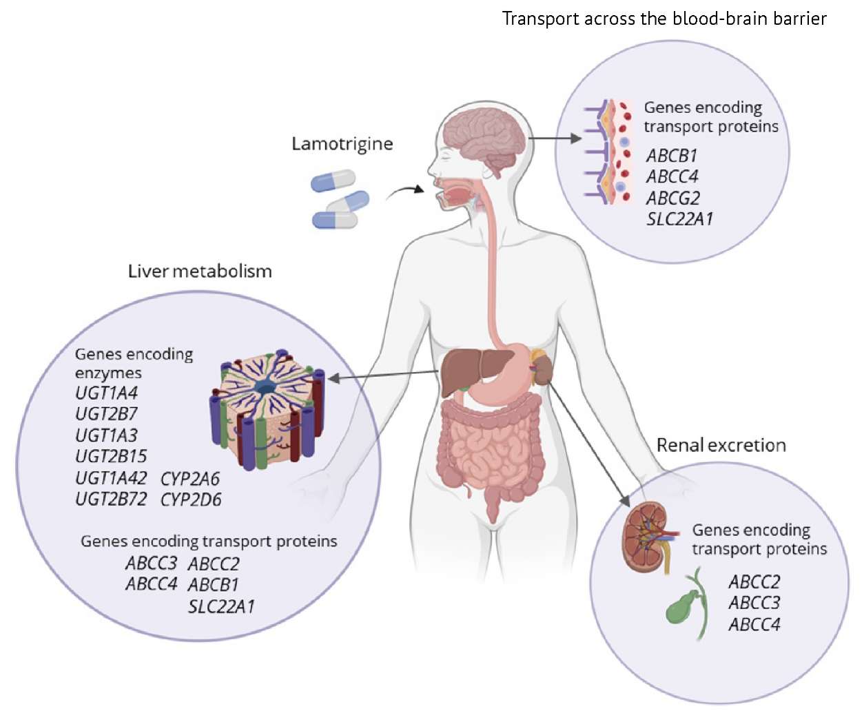
The figure was prepared by the authors
Fig. 2. Genes responsible for lamotrigine metabolism and transport in the human body
The main LTG metabolites resulting from glucuronidation (Table 2) are inactive (neutral) and excreted by the kidneys [61]. However, some studies demonstrate that increased LTG glucuronides in the urine correlate with a higher level of its active metabolites in blood plasma. Examination of LTG glucuronides in urine samples is a non-invasive and promising strategy for safety assessment of LTG therapy that can be implemented using gas chromatography-mass spectrometry or micellar electrokinetic capillary chromatography [61].
Increased blood levels of active LTG metabolites resulting from P-oxidation (Table 2) are associated with neurotoxic, dermatotoxic, hepatotoxic, and cardiotoxic ADRs [28]. However, the exact production mechanism and the toxicity of these metabolites based on their levels in the blood, brain (LTG target organ), and other tissues warrant a further study.
LTG neurotoxicity is rare and manifests itself as ataxia [62], dizziness, and diplopia, especially in newborns, infants under one year [63], women of reproductive age [7], and older patients [64]. However, this ADR can lead to more severe problems including epileptic seizures (even in individuals without a history of seizures) [65–68]; optic neuritis with a sudden loss of vision; altered state of consciousness (Glasgow coma scale ≤8); severe arterial hypotension; torsade de pointes ventricular tachycardia; and cardiac arrest (fatal cases reported for LTG 4 g and 7.5 g) [68]. LTG neurotoxicity also depends on an individual genotype, including polymorphisms in genes encoding key metabolic enzymes and transporter proteins involved in LTG efflux from the brain into blood (phenotypes: slow and intermediate metabolisers and / or slow and intermediate transporters) [66]. These genetically determined interindividual differences in LTG serum and brain levels may be critical for neurotoxic ADRs, since delayed BBB clearance may cause accumulation of toxic LTG metabolites in the brain during long-term high-dose therapy, as well as in postpartum women with breastfed infants [7].
Overall, a personalised multi-omics approach (pharmacogenomics + pharmacometabolomics) to LTG therapy reflects current clinical and laboratory diagnostic trends in neurology and psychiatry [66][69]. The risk of neurotoxicity is higher in patients who are slow metabolisers and/or slow transporters (homozygous carriers of non-functional polymorphisms in the genes of LTG glucuronidation and / or P-oxidation isoenzymes (Table 3) and/or genes encoding key LTG transporter proteins (Table 4), respectively) compared to normal metabolisers and normal transporters (homozygous carriers of fully functional polymorphisms in genes encoding key LTG isoenzymes and transporter proteins, respectively). However, the prevalence of these phenotypes among patients taking LTG is unknown, since studies using the multi-omics approach are not yet available.
To assess the risk of LTG-induced ADRs, a clinical evaluation оf the patient's condition [70] is recommended, alongside with 1) therapeutic drug monitoring (TDM) of LTG (blood, hair, saliva, breast milk) [70]; 2) assay of active (potentially toxic) LTG metabolites in biological samples (blood, saliva, hair) using gas-liquid chromatography; 3) pharmacogenetic testing (PGx) of carriers of non-functional polymorphisms in genes encoding key transporter proteins and enzymes involved in LTG metabolism [66][71].
TDM ensures optimal LTG dose adjustment. Minimally invasive sample collection strategies for TDM (dried blood spot [70] or plasma [72], saliva, or hair [73][74]) are promising for wide clinical adoption, allowing self-sampling, room temperature storage and transport. In certain clinical conditions (pregnancy, neonatal period, ageing, resistance, and toxicity), measuring LTG and active metabolites in plasma is reliable and sufficient. However, liquid chromatography-tandem mass spectrometry of blood, saliva, and hair is promising for safety monitoring and early toxicity diagnosis. LTG hair analysis can also be used to assess medication history and long-term adherence of a patient. LTG saliva and plasma concentrations correlate strongly (r=0.82, p<0.001); LTG hair concentration correlates with plasma concentrations (r=0.53, p<0.001) and daily dose (r=0.23, p=0.024) [74].
Predictive (pre-reactive) PGx will help assess the risk of LTG and its active metabolites increasing to toxic levels in order to exclude homozygous (primarily) or heterozygous non-functional polymorphisms in genes responsible for its metabolism and transport [66][71][75][76]. For slow and intermediate metabolisers and / or transporters, it is recommended to avoid LTG prescription or reduce the average daily dose by 50% and 25%, respectively; to conduct TDM once every 3 and 6 months, respectively. If technically feasible, measurement of active LTG metabolites in biological fluids (plasma or serum, saliva, urine) is important. In case of potentially life-threatening toxicity symptoms, extracorporeal LTG elimination (haemodialysis, plasmapheresis) may be considered [77][78].
The analysed pharmacogenomic and pharmacometabolic data on LTG metabolism, transport, and elimination show that the main active (potentially toxic) metabolites are LTG-N-oxide and LTG-N-methyl; their high blood and brain (target organ) concentrations can cause neurotoxicity. The high-risk group includes newborns and infants, pregnant women, older population, and patients with unfavourable pharmacogenetic profiles (intermediate and poor metabolisers) who are heterozygous and homozygous carriers of non-functional polymorphisms in genes encoding key glucuronidation enzymes (UGT1A4, UGT2B7, UGT1A3) and, to a lesser extent, P-oxidation (CYP2D6, CYP2A6) of LTG.
Reduced LTG efflux transport from brain to blood in patients with unfavourable pharmacogenetic transporter profiles (intermediate transporters and poor transporters - heterozygous and homozygous carriers of non-functional polymorphisms in genes encoding key LTG transporter proteins ABCC3, ABCC4, ABCC2, ABCB1, and SLC22A1) also significantly contributes to increased risk of severe LTG-induced neurotoxic ADRs.
The above approach using pharmacogenomic and pharmacometabolic data will enable a more accurate prediction of individual responses to LTG and reduce the risk of toxic ADRs. Advanced multi-omics technologies and artificial intelligence may facilitate the implementation of multi-omics phenotyping in real clinical practice, considering such factors as pharmacogenomics, pharmaco-metabolomics, environmental factors, comorbidities, and patient lifestyle for personalising psychopharmacotherapy.
1. Lamotrigine. https://go.drugbank.com/drugs/DB00555
2. https://www.prisma-statement.org/prisma-2020
3. Lamotrigine. https://go.drugbank.com/drugs/DB00555
4. Lamotrigine. https://go.drugbank.com/drugs/DB00555
5. Lamotrigine. https://go.drugbank.com/drugs/DB00555
6. Human Metabolome Database. https://hmdb.ca/
7. https://pubchem.ncbi.nlm.nih.gov/
8. https://www.pharmgkb.org/chemical/PA166170365
9. https://www.ncbi.nlm.nih.gov/snp/
10. Lamotrigine. https://go.drugbank.com/drugs/DB00555
11. Online Mendelian Inheritance in Man®. https://www.omim.org/
12. Ibid.
1. Panebianco M, Bresnahan R, Marson AG. Lamotrigine add-on therapy for drug-resistant focal epilepsy. Cochrane Database Syst Rev. 2023;12(12):CD001909. https://doi.org/10.1002/14651858.CD001909.pub4
2. Campbell R, Beall J. Pharmacogenomics of lamotrigine: A possible link to serious cutaneous adverse reactions. Ment Health Clin. 2015;5(2):78–81. https://doi.org/10.9740/mhc.2015.03.078
3. Shilkina OS, Zobova SN, Domoratskaya EA, Dmitrenko DV. Clinical and genetic characteristics of juvenile myoclonic epilepsy. Personalized Psychiatry and Neurology. 2021;1(2):95–105. https://doi.org/10.52667/2712-9179-2021-1-2-95-105
4. Besag FMC, Vasey MJ, Sharma AN, Lam ICH. Efficacy and safety of lamotrigine in the treatment of bipolar disorder across the lifespan: A systematic review. Ther Adv Psychopharmacol. 2021;11:20451253211045870. https://doi.org/10.1177/20451253211045870
5. Hashimoto Y, Kotake K, Watanabe N, et al. Lamotrigine in the maintenance treatment of bipolar disorder. Cochrane Database Syst Rev. 2021;9(9):CD013575. https://doi.org/10.1002/14651858.CD013575.pub2
6. Katayama Y, Terao T, Kamei K, et al. Therapeutic window of lamotrigine for mood disorders: A naturalistic retrospective study. Pharmacopsychiatry. 2014;47(3):111–4. https://doi.org/10.1055/s-0034-1375618
7. Dmitrenko DV, Shnayder NA, Egorova AT. Epilepsy and pregnancy. Moscow: GEOTAR-Media; 2022 (In Russ.). https://doi.org/10.33029/9704-6359-8-EPI-2022-1-296
8. Goo Y, der Nederlanden AM, Bleasel A, et al. Dose monitoring of lamotrigine monotherapy in pregnancy: Are pregnant women with epilepsy currently optimally managed? A systematic review. Ther Drug Monit. 2024;46(2):181–94. https://doi.org/10.1097/FTD.0000000000001186
9. Hope OA, Harris KM. Management of epilepsy during pregnancy and lactation. BMJ. 2023;382:e074630. https://doi.org/10.1136/bmj-2022-074630
10. Anderson GD. Pregnancy-induced changes in pharmacokinetics: A mechanistic-based approach. Clin Pharmacokinet. 2005;44(10):989–1008. https://doi.org/10.2165/00003088-200544100-00001
11. Viale L, Allotey J, Cheong-See F, et al. Epilepsy in pregnancy and reproductive outcomes: A systematic review and meta-analysis. Lancet. 2015;386(10006):1845–52. https://doi.org/1016/S0140-6736(15)00045-8
12. Berry-Noronha A, Manoleehakul P, Rottler A, et al. Risk of adverse pregnancy outcomes associated with antiseizure medications and their indications: A systematic review and meta-analysis. Neurology. 2025;104(3):e210233. https://doi.org/10.1212/WNL.0000000000210233
13. Qian Y, Wu W, Ke C, et al. Toward precision dosing of lamotrigine during pregnancy: Physiologically based pharmacokinetic modeling and simulation. CPT Pharmacometrics Syst Pharmacol. 2025;14(5):929–40. https://doi.org/10.1002/psp4.70007
14. Yan R, Tuo J, Tai Z, et al. Management of anti-seizure medications in lactating women with epilepsy. Front Neurol. 2022;13:1005890. https://doi.org/10.3389/fneur.2022.1005890
15. Hasser C, Ameresekere M, Girgis C, et al. Striking the balance: Bipolar disorder in the perinatal period. Focus (Am Psychiatr Publ). 2024;22(1):3–15. https://doi.org/10.1176/appi.focus.20230020
16. Nordmo E, Aronsen L, Wasland K, et al. Severe apnea in an infant exposed to lamotrigine in breast milk. Ann Pharmacother. 2009;43(11):1893–97. https://doi.org/10.1345/aph.1M254
17. Clark CT, Klein AM, Perel JM, et al. Lamotrigine dosing for pregnant patients with bipolar disorder. Am J Psychiatry. 2013;170(11):1240–7. https://doi.org/10.1176/appi.ajp.2013.13010006
18. Nucera B, Brigo F, Trinka E, Kalss G. Treatment and care of women with epilepsy before, during, and after pregnancy: A practical guide. Ther Adv Neurol Disord. 2022;15:17562864221101687. https://doi.org/10.1177/17562864221101687
19. Arfman IJ, Wammes-van der Heijden EA, Ter Horst PGJ, et al. Therapeutic drug monitoring of antiepileptic drugs in women with epilepsy before, during, and after pregnancy. Clin Pharmacokinet. 2020;59(4):427–45. https://doi.org/10.1007/s40262-019-00845-2
20. Shawahna R, Zaid L. Concentrations of antiseizure medications in breast milk of lactating women with epilepsy: A systematic review with qualitative synthesis. Seizure. 2022;98:57–70. https://doi.org/10.1016/j.seizure.2022.03.017
21. Babu M, Snyder M. Multi-omics profiling for health. Mol Cell Proteomics. 2023;22(6):100561. https://doi.org/10.1016/j.mcpro.2023.100561
22. Zhou Y, Wang X, Li H, et al. Polymorphisms of ABCG2, ABCB1 and HNF4α are associated with lamotrigine trough concentrations in epilepsy patients. Drug Metab Pharmacokinet. 2015;30(4):282–7. https://doi.org/10.1016/j.dmpk.2015.05.002
23. Römermann K, Helmer R, Löscher W. The antiepileptic drug lamotrigine is a substrate of mouse and human breast cancer resistance protein (ABCG2). Neuropharmacology. 2015;93:7–14. https://doi.org/10.1016/j.neuropharm.2015.01.015
24. Azhigova AM, Broutian AG, Vlasov PN. The pharmacogenomics of lamotrigine (a literature review). Annals of Clinical and Experimental Neurology. 2021;15(2):59–72 (In Russ.). https://doi.org/10.25692/ACEN.2021.2.8
25. Zelcer N, van de Wetering K, de Waart R, et al. Mice lacking Mrp3 (Abcc3) have normal bile salt transport, but altered hepatic transport of endogenous glucuronides. J Hepatol. 2006;44(4):768–75. https://doi.org/10.1016/j.jhep.2005.07.022
26. Dickens D, Owen A, Alfirevic A, et al. Lamotrigine is a substrate for OCT1 in brain endothelial cells. Biochem Pharmacol. 2012;83(6):805–14. https://doi.org/10.1016/j.bcp.2011.12.032
27. Vázquez M, Fagiolino P. The role of efflux transporters and metabolizing enzymes in brain and peripheral organs to explain drug-resistant epilepsy. Epilepsia Open. 2022;1(l1):S47–S58. https://doi.org/10.1002/epi4.12542
28. Mitra-Ghosh T, Callisto SP, Lamba JK, et al. PharmGKB summary: Lamotrigine pathway, pharmacokinetics and pharmacodynamics. Pharmacogenet Genomics. 2020;30(4):81–90. https://doi.org/10.1097/FPC.0000000000000397
29. Ademuwagun IA, Rotimi SO, Syrbe S, et al. Voltage gated sodium channel genes in epilepsy: Mutations, functional studies, and treatment dimensions. Front Neurol. 2021;12:600050. https://doi.org/10.3389/fneur.2021.600050
30. Pejanovic-Skobic N, Markovic I, Bozina N, Basic S. Lack of association of SCN2A rs17183814 polymorphism with the efficacy of lamotrigine monotherapy in patients with focal epilepsy from Herzegovina area, Bosnia and Herzegovina. Epilepsy Res. 2019;158:106221. https://doi.org/10.1016/j.eplepsyres.2019.106221
31. Glauser TA, Holland K, O’Brien VP, et al. Pharmacogenetics of antiepileptic drug efficacy in childhood absence epilepsy. Ann Neurol. 2017;81(3):444–53. https://doi.org/10.1002/ana.24886
32. Robakis TK, Holtzman J, Stemmle PG, et al. Lamotrigine and GABAA receptor modulators interact with menstrual cycle phase and oral contraceptives to regulate mood in women with bipolar disorder. J Affect Disord. 2015;175:108–15. https://doi.org/10.1016/j.jad.2014.12.040
33. Costa B, Vale N. Understanding lamotrigine’s role in the CNS and possible future evolution. Int J Mol Sci. 2023;24(7):6050. https://doi.org/10.3390/ijms24076050
34. Paraskevas GP, Triantafyllou NI, Kapaki E, et al. Add-on lamotrigine treatment and plasma glutamate levels in epilepsy: Relation to treatment response. Epilepsy Res. 2006;70(2–3):184–9. https://doi.org/10.1016/j.eplepsyres.2006.05.004
35. Zhao T, Zhang HL, Feng J, et al. Impact of UGT1A4 and UGT2B7 polymorphisms on lamotrigine plasma concentration in patients with bipolar disorder. Pharmacogenet Genomics. 2024;34(8):261–7. https://doi.org/10.1097/FPC.0000000000000543
36. Petrenaite V, Öhman I, Jantzen FPT, Ekström L. Effect of UGT1A4, UGT2B7, UGT2B15, UGT2B17 and ABC1B polymorphisms on lamotrigine metabolism in Danish patients. Epilepsy Res. 2022;182:106897. https://doi.org/10.1016/j.eplepsyres.2022.106897
37. Kim SC, Kim MG. Meta-analysis of the Influence of UGT genetic polymorphisms on lamotrigine concentration. Basic Clin Pharmacol Toxicol. 2019;124(2):163–9. https://doi.org/10.1111/bcpt.13120
38. Liu L, Zhao L, Wang Q, et al. Influence of valproic acid concentration and polymorphism of UGT1A4*3, UGT2B7 –161C > T and UGT2B7*2 on serum concentration of lamotrigine in Chinese epileptic children. Eur J Clin Pharmacol. 2015;71(11):1341–7. https://doi.org/10.1007/s00228-015-1925-9
39. Wang Q, Zhao L, Liang M, et al. Effects of UGT2B7 genetic polymorphisms on serum concentrations of valproic acid in Chinese children with epilepsy comedicated with lamotrigine. Ther Drug Monit. 2016;38(3):343–9. https://doi.org/10.1097/FTD.0000000000000271
40. Edavana VK, Dhakal IB, Williams S, et al. Potential role of UGT1A4 promoter SNPs in anastrozole pharmacogenomics. Drug Metab Dispos. 2013;41(4):870–7. https://doi.org/10.1124/dmd.112.048157
41. Reimers A, Sjursen W, Helde G, Brodtkorb E. Frequencies of UGT1A4*2 (P24T) and *3 (L48V) and their effects on serum concentrations of lamotrigine. Eur J Drug Metab Pharmacokinet. 2016;41(2):149–55. https://doi.org/10.1007/s13318-014-0247-0
42. Petrenaite V, Öhman I, Ekström L, et al. UGT polymorphisms and lamotrigine clearance during pregnancy. Epilepsy Res. 2018;140:199–208. https://doi.org/10.1016/j.eplepsyres.2018.01.011
43. Wang Z, Wong T, Hashizume T, et al. Human UGT1A4 and UGT1A3 conjugate 25-hydroxyvitamin D3: metabolite structure, kinetics, inducibility, and interindividual variability. Endocrinology. 2014;155(6):2052–63. https://doi.org/10.1210/en.2013-2013
44. Mimura Y, Maruo Y, Ohta Y, et al. Effect of common exon variant (p.P364L) on drug glucuronidation by the human UDP-glucuronosyltransferase 1 family. Basic Clin Pharmacol Toxicol. 2011;109(6):486–93. https://doi.org/10.1111/j.1742-7843.2011.00754.x
45. Blanca Sánchez M, Herranz JL, Leno C, et al. UGT2B7 –161C>T polymorphism is associated with lamotrigine concentration-to-dose ratio in a multivariate study. Ther Drug Monit. 2010;32(2):177–84. https://doi.org/10.1097/FTD.0b013e3181ceecc6
46. Singkham N, Towanabut S, Lertkachatarn S, Punyawudho B. Influence of the UGT2B7 –161C>T polymorphism on the population pharmacokinetics of lamotrigine in Thai patients. Eur J Clin Pharmacol. 2013;69(6):1285–91. https://doi.org/10.1007/s00228-012-1449-5
47. Milosheska D, Lorber B, Vovk T, et al. Pharmacokinetics of lamotrigine and its metabolite N-2-glucuronide: Influence of polymorphism of UDP-glucuronosyltransferases and drug transporters. Br J Clin Pharmacol. 2016;82(2):399–411. https://doi.org/10.1111/bcp.12984
48. Provenzani A, Labbozzetta M, Notarbartolo M, et al. Rash and multiorgan dysfunction following lamotrigine: could genetic be involved? Int J Clin Pharm. 2015;37(5):682–6. https://doi.org/10.1007/s11096-015-0158-4
49. Rowland A, Elliot DJ, Williams JA, et al. In vitro characterization of lamotrigine N2-glucuronidation and the lamotrigine-valproic acid interaction. Drug Metab Dispos. 2006;34(6):1055–62. https://doi.org/10.1124/dmd.106.009340
50. Lu W, Uetrecht JP. Possible bioactivation pathways of lamotrigine. Drug Metab Dispos. 2007;35(7):1050–6. https://doi.org/10.1124/dmd.107.015271
51. Chen H, Grover S, Yu L, et al. Bioactivation of lamotrigine in vivo in rat and in vitro in human liver microsomes, hepatocytes, and epidermal keratinocytes: characterization of thioether conjugates by liquid chromatography/mass spectrometry and high field nuclear magnetic resonance spectroscopy. Chem Res Toxicol. 2010;23(1):159–70. https://doi.org/10.1021/tx9003243
52. Nasyrova RF, Shnayder NA, Osipova SM, et al. Genetic predictors of antipsychotic efflux impairment via blood-brain barrier: Role of transport proteins. Genes (Basel). 2023;14(5):1085. https://doi.org/10.3390/genes14051085
53. Klarica Domjanović I, Lovrić M, Trkulja V, et al. Interaction between ABCG2 421C>A polymorphism and valproate in their effects on steady-state disposition of lamotrigine in adults with epilepsy. Br J Clin Pharmacol. 2018;84(9):2106–19. https://doi.org/10.1111/bcp.13646
54. Shen CH, Zhang YX, Lu RY, et al. Specific OCT1 and ABCG2 polymorphisms are associated with lamotrigine concentrations in Chinese patients with epilepsy. Epilepsy Res. 2016;127:186–90. https://doi.org/10.1016/j.eplepsyres.2016.09.004
55. Qu J, Zhou BT, Yin JY, et al. ABCC2 polymorphisms and haplotype are associated with drug resistance in Chinese epileptic patients. CNS Neurosci Ther. 2012;18(8):647–51. https://doi.org/10.1111/j.1755-5949.2012.00336.x
56. Sasaki T, Hirota T, Ryokai Y, et al. Systematic screening of human ABCC3 polymorphisms and their effects on MRP3 expression and function. Drug Metab Pharmacokinet. 2011;26(4):374–86. https://doi.org/10.2133/dmpk.dmpk-10-rg-103
57. Nies AT, Jedlitschky G, König J, et al. Expression and immunolocalization of the multidrug resistance proteins, MRP1-MRP6 (ABCC1-ABCC6), in human brain. Neuroscience. 2004;129(2):349–60. https://doi.org/10.1016/j.neuroscience.2004.07.051
58. Rius M, Nies AT, Hummel-Eisenbeiss J, et al. Cotransport of reduced glutathione with bile salts by MRP4 (ABCC4) localized to the basolateral hepatocyte membrane. Hepatology. 2003;38(2):374–84. https://doi.org/10.1053/jhep.2003.50331
59. Berthier J, Arnion H, Saint-Marcoux F, Picard N. Multidrug resistance-associated protein 4 in pharmacology: Overview of its contribution to pharmacokinetics, pharmacodynamics and pharmacogenetics. Life Sci. 2019;231:116540. https://doi.org/10.1016/j.lfs.2019.06.015
60. Bazhenova A, Mironov K, Kravchenko A, Akimkin V. Pharmacogenetic effects of single nucleotide polymorphisms commonly associated with antiretroviral therapy metabolism. HIV Infection and Immunosuppressive Disorders. 2022;14(3):65–76 (In Russ.). https://doi.org/10.22328/2077-9828-2022-14-3-65-76
61. Pucci V, Bugamelli F, Baccini C, Raggi MA. Analysis of lamotrigine and its metabolites in human plasma and urine by micellar electrokinetic capillary chromatography. Electrophoresis. 2005;26(4–5):935–42. https://doi.org/10.1002/elps.200410208
62. Wood KE, Palmer KL, Krasowski MD. Correlation of elevated lamotrigine and levetiracetam serum/plasma levels with toxicity: A long-term retrospective review at an academic medical center. Toxicol Rep. 2021;8:1592–8. https://doi.org/10.1016/j.toxrep.2021.08.005
63. Close BR, Banks CJ. Seizures secondary to lamotrigine toxicity in a two-year-old. Ann Pharmacother. 2010;44(6):1112–5. https://doi.org/10.1345/aph.1M617
64. Moore PW, Donovan JW, Burkhart KK, Haggerty D. A case series of patients with lamotrigine toxicity at one center from 2003 to 2012. Clin Toxicol (Phila). 2013;51(7):545–9. https://doi.org/10.3109/15563650.2013.818685
65. Chiew AL, Isbister GK, Nguyen K, et al. Clinical effects of acute lamotrigine overdose (ATOM-10). Clin Toxicol (Phila). 2025;63(5):310–6. https://doi.org/10.1080/15563650.2025.2471906
66. Nasyrova RF, Neznanov NG, eds. Clinical psychopharmacogenetics. St. Petersburg: DEAN; 2019 (In Russ.). EDN: QCOSIL
67. Alyahya B, Friesen M, Nauche B, Laliberté M. Acute lamotrigine overdose: a systematic review of published adult and pediatric cases. Clin Toxicol (Phila). 2018;56(2):81–9. https://doi.org/10.1080/15563650.2017.1370096
68. Chou M, Lai L, Neveu M, Ritchie A. Toxic optic neuropathy associated with lamotrigine and levetiracetam dual therapy. BMJ Case Rep. 2024;17(3):e256961. https://doi.org/10.1136/bcr-2023-256961
69. Shnayder NA, Grechkina VV, Arkhipov VV, Nasyrova RF. Pharmacogenetics-informed pharmacometabolomics as an innovative approach to assessing the safety and risk of pharmacotherapy with valproic acid. Safety and Risk of Pharmacotherapy. 2023;11(4):450–62 (In Russ.). https://doi.org/10.30895/2312-7821-2023-386
70. Min KL, Ryu JY, Chang MJ. Development and clinical applications of the dried blood spot method for therapeutic drug monitoring of anti-epileptic drugs. Basic Clin Pharmacol Toxicol. 2019;125(3):215–36. https://doi.org/10.1111/bcpt.13269
71. Milosavljevic F, Manojlovic M, Matkovic L, et al. Pharmacogenetic variants and plasma concentrations of antiseizure drugs: A syste matic review and meta-analysis. JAMA Netw Open. 2024;7(8):e2425593. https://doi.org/10.1001/jamanetworkopen.2024.25593
72. Cao H, Jiang Y, Sun Q, et al. Simultaneous monitoring of seven antiepileptic drugs by dried blood spot and dried plasma spot sampling: method validation and clinical application of a LC-MS/MS-based technique. J Pharm Biomed Anal. 2024;243:116099. https://doi.org/10.1016/j.jpba.2024.116099
73. Pelcová M, Ďurčová V, Šmak P, et al. Non-invasive therapeutic drug monitoring: LC-MS validation for lamotrigine quantification in dried blood spot and oral fluid/saliva. J Pharm Biomed Anal. 2025;262:116877. https://doi.org/10.1016/j.jpba.2025.116877
74. Kuczynska J, Karas-Ruszczyk K, Zakrzewska A, et al. Comparison of plasma, saliva, and hair lamotrigine concentrations. Clin Biochem. 2019;74:24–30. https://doi.org/10.1016/j.clinbiochem.2019.09.009
75. Shnayder NA, Grechkina VV, Arkhipov VV, Nasyrova RF. The role of pharmacogenetic testing in assessing the risk and safety of valproate use: An ethnic aspect (review). Safety and Risk of Pharmacotherapy. 2024;12(2):14–36 (In Russ.). https://doi.org/10.30895/2312-7821-2024-12-2-14-36
76. Amaro-Álvarez L, Cordero-Ramos J, Calleja-Hernández MÁ. Exploring the impact of pharmacogenetics on personalized medicine: A systematic review. Farm Hosp. 2024;48(6):299–309. https://doi.org/10.1016/j.farma.2023.12.004
77. Gosselin S, Ghannoum M, Hoffman RS. Hemodialysis for lamotrigine poisoning. Am J Emerg Med. 2020;38(2):403–4. https://doi.org/10.1016/j.ajem.2019.158385
78. Agrawal A, Nogar JN, Koenig S. Management of lamotrigine overdose using hemodialysis. Am J Emerg Med. 2019;37(8):1603.e1–1603.e2. https://doi.org/10.1016/j.ajem.2019.05.02
Natalia А. Shnayder, Dr. Sci. (Med.), Professor
3 Bekhterev St., St Petersburg 192019;
1 Partizan Zheleznyak St., Krasnoyarsk 660022
Violetta V. Bader
3 Bekhterev St., St Petersburg 192019;
9 Obvodny Canal Emb., St. Petersburg 191167
Regina F. Nasyrova, Dr. Sci. (Med.)
3 Bekhterev St., St Petersburg 192019;
92 Lenin Ave., Tula 300012
Mikhail Ya. Kissin, Dr. Sci. (Med.), Professor
9 Obvodny Canal Emb., St. Petersburg 191167;
6–8, Lev Tolstoy St., St Petersburg 197022
Shnayder N.A., Bader V.V., Nasyrova R.F., Kissin M.Ya. Multi-Omics in Predicting Lamotrigine Neurotoxicity: Current Opportunities (Review). Safety and Risk of Pharmacotherapy. 2026;14(1):51-65. https://doi.org/10.30895/2312-7821-2025-498
 Email this article (Login required)
Email the author (Login required)
Email this article (Login required)
Email the author (Login required) Publisher: Scientific Centre for Expert Evaluation of Medicinal Products
Processing of personal data





